FieldStrength MRI articles
User experiences - July 2015
"The MultiVane XD technique is a savior in clinical diagnostic quality in our most challenging patients"
Ben Kennedy, chief MRI technologist at Qscan Radiology Clinic (Brisbane, Australia) explains how MultiVane XD helps them in dealing with respiratory motion in MRI of shoulder and elbow.

Ben Kennedy, chief MRI technologist at Qscan Radiology Clinic in Brisbane, Australia
“MultiVane XD has been easy to implement and allows me to stick to my basic sequence optimization steps for obtaining high image quality.”
When motion affects MRI of upper limbs
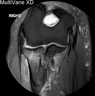
“As upper limb joints are in direct connection to the chest, MR imaging of these joints is susceptible to breathing artifacts, particularly when scanning in axial orientation. Routine MR imaging of the shoulder and elbow regions benefits greatly from motion reduction with MultiVane XD. In our experience, particularly the first sequence of an exam is likely to suffer from respiratory motion as patients are settling into the magnet environment. However, some patients naturally have a lot of respiratory motion, for instance athletes carrying a higher muscle bulk or obese patients. For such cases we have optimized MultiVane XD for all directions with all TSE sequences of the shoulder and elbow.”
Integrating MultiVane XD in the exam
“MultiVane XD has been easy to implement and allows me to stick to my basic sequence optimization steps for obtaining high image quality. Compared to the previous version of MultiVane that we used, we find MultiVane XD providing some significant improvements [1] in flexibility and confidence in our imaging outcomes.” “Using MultiVane XD our non-fat saturated sequences show high definition of the trabecular pattern, which is important in high detail musculoskeletal imaging.”

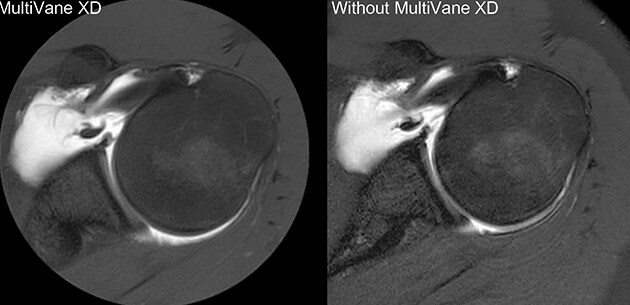
Comparison of axial fat suppressed PD-weighted imaging with and without MultiVane XD in the left shoulder demonstrates that the imaging with MultiVane XD provides excellent image quality, even in presence of motion.

Imaging with MultiVane XD provides excellent image quality, even in presence of motion.
Different sequences, different approaches
“For PD and T2 fat suppressed imaging, I prefer the asymmetric k-space sampling that gives me great flexibility in tailoring each sequence to enhance image quality and speed. It allows me to increase SNR without compromising voxel size, so that I maintain the same high resolution, and maintain the preferred TE position within the shot length for the desired image quality. Using asymmetric sampling also helps maximize image contrast, which is paramount in musculoskeletal imaging.” “For T1 and T1 fat suppressed imaging, we can still use low-high k-space sampling and stick to the same maximum shot length rule as in our standard TSE version. We are using multiple shots per blade to widen the width of the phase sampling blade. At 3.0T, we have the ability to keep TE spacing much shorter, which requires a smaller water fat shift, whilst maintaining good SNR thanks to dStream.”
Final considerations
“For blade percentage, I start at minimum 200% and reduce any multiple NSA to 1, as multiple NSA will overlay multiple corrected image data samples, which may not correct the same. By choosing the MultiVane percentage as NSA (e.g. 4 NSA would be the same as 400%) for keeping SNR equivalent, all data will be corrected to the same image reduction. For data at minimum 200%, which started at 1 NSA, dS SENSE can be used for reducing the scan time back where it started.” “After optimizing the contrast parameters, it is important to recognize that often a slightly larger field of view is needed to cover the desired anatomy when converting from a square or rectangular FOV to a circular FOV in order. Increasing FOV can also be a simple solution to get rid of a wrap artifact from a larger size patient.”
“The ability to use dS SENSE can be a great benefit for speed.”
Concluding remarks
“The ability to use dS SENSE can be a great benefit for speed where required. Its sampling pattern preserves SNR even in higher SENSE reduction values. In other sequences, we take advantage of the SENSE unwrapping algorithm at the reduction value of 1, which I commonly leave on.” “Thanks to the homogeneous wide bore magnet of Ingenia, standard SPAIR and SPIR fat saturation is still very robust on these protocols, which use supine patient positioning with arm by the side.” “The MultiVane XD technique is a savior in clinical diagnostic quality in our most challenging patients. You can have the best equipment available but capturing a moving target requires this next step of innovation.”

Reference [1] Pipe et al., Magn Reson Med. 2014;72(2):430-7 Results obtained at this facility may not be typical for other facilities

Related information
More from FieldStrength
