FieldStrength MRI magazine
User experiences - July 2020
Ingenia Ambition brings consistent image quality, high resolution, and fantastic patient comfort to Altona Children’s Hospital
For pediatric patients undergoing MRI at Altona Children’s Hospital, the staff does what they can to get a confident diagnosis right away, in the first MRI exam. While the room lighting and the immersive in-bore audiovisual experience help their patients feel at ease, the experienced staff takes advantage of the features and capabilities of their recently acquired Ingenia Ambition scanner. The excellent image quality and the flexibility to adapt the exam to the findings, allows the staff to serve their very diverse patient group, from tiny newborns and toddlers to teenagers, very well.
“Essential to us are the consistent quality and resolution of the images, for every sequence”
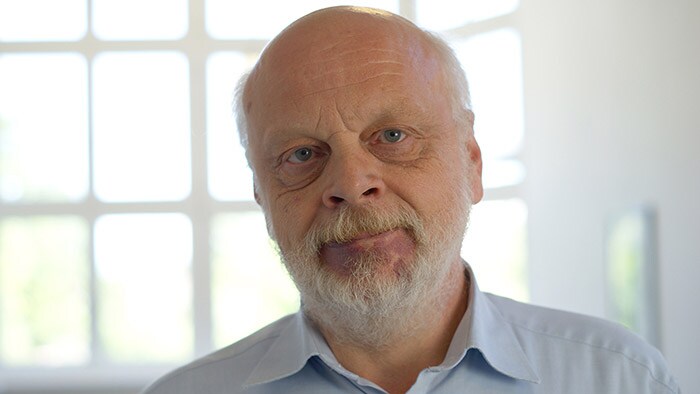
Carl-Martin Junge, MD Chief Radiologist, General and Pediatric Radiology at Altona Children’s Hospital. He has more than 25 years of experience with MRI.
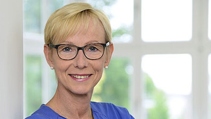
Frauke Meinken MRI lead technologist at Altona Children’s Hospital
"With Ambition, we can perform high quality abdominal scans. As a result, the number of abdominal scans has increased substantially compared to our practice with our old system.”
Consistent image quality as top priority for children of every age and size
Dr. Carl-Martin Junge is the Chief Radiologist at the Pediatric Radiology Department. Since November 2019, the department has been using the Philips Ingenia Ambition 1.5T MRI system. Dr. Junge explains that the large range in age and size of pediatric patients, requires a broad set of MRI examination protocols. His first priority for an MRI scanner is good and consistent image quality, so that he is able to make confident diagnoses in even the smallest and most difficult pediatric patients. In addition, their way of working requires an MRI scanner that offers a high degree of flexibility to allow tailoring of their imaging on the fly when initial findings give them a reason to do so. From day one of operation, the Ingenia Ambition has impressed Dr. Junge and his team with its powerful performance.
The Pediatric Radiology Department of Altona Children’s Hospital in Hamburg, Germany, performs diagnostic imaging in pediatric patients, from newborns to patients of about 18 years of age, specializing in orthopedic, neurological, and gastroenterological imaging.
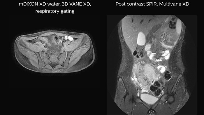
Crohn’s disease in the terminal ileum A large abscess is visible near the terminal ileum, in the middle of the coronal image.
Overcoming challenges associated with pediatric imaging
Dr. Junge estimates that per day, on average two or three patients out of ten would be scanned with sedation. MRI lead technologist Ms. Meinken adds that she has been happily surprised that the need for sedation was not affected when switching from an open system to a bore system – thanks to the fantastic in-bore and Ambient features. Another important factor is that many pediatric patients are still undiagnosed at the time of presentation, and the desire is to find a diagnosis immediately in the first exam, in order to limit or avoid the need for follow-up exams. As a consequence, the team needs flexibility and time during the examination.
Dr. Junge points out how scanning pediatric patients is more difficult than scanning adults. In addition to the small size, age range and often complex diagnoses, he mentions the challenges with keeping younger patients still in the scanner, during scans that often require 30 minutes or more. “For children from zero to about five years old we use sedation to successfully carry out the examination. For patients from five to about 12-13 years, good coaching and the wonderful Ambient and in-bore features help keep the children calm in the machine and allow for good imaging. It’s a very good advantage compared to other systems, I think. Older children can be treated similar to adults. Of course, motion-robust scan techniques help to address these challenges for all ages.”
“The in-bore features are key to successful examinations, especially for children”
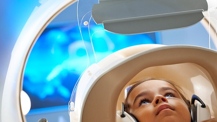
Ms. Meinken notes that the Ambient Experience also makes the atmosphere of the scanning room much more pleasant for the staff. “It’s nice for the technologists to work in this friendly-feeling room. It feels much better than working in a dull, colorless room, as we did in the past,” she says.
Patients and parents benefit from the many features designed for their comfort
“And the parents love it too,” says Dr. Junge. “In addition, we can use noise reduction to make the scan more comfortable. Patients with spondylolisthesis or other painful afflictions particularly appreciate the special thick, soft mattress, which helps make their examinations much more comfortable. Many of these patients say it’s fantastic, and they were never lying so comfortable in an MRI system before. I absolutely think that Ambition improves patient satisfaction.”
Both Dr. Junge and Ms. Meinken value the patient-friendly attributes of the Ingenia Ambition. “The in-bore features, including music and videos to entertain the children during the scanning, are very useful,” says Ms. Meinken. “The children really love this, it helps us persuade many of them. Without this, young children often do not want to enter the scanner, but with the films, music and color, they get interested and willing to undergo the scan. And we can take our time with the examinations, because the patients are distracted and at ease. I think, the in-bore features are key to successful examinations, especially for children.”
“These capabilities also help the patients remain motionless inside the scanner, which allows us to obtain images with excellent quality,” says Dr. Junge. “I think the in-bore Connect capability is one of the best highlights of this system.”
Ms. Meinken notes that the Ambient Experience also makes the atmosphere of the scanning room much more pleasant for the staff. “It’s nice for the technologists to work in this friendly-feeling room. It feels much better than working in a dull, colorless room, as we did in the past,” she says.
“It’s nice for the technologists to work in this friendly-feeling room.”
Achieving consistent high quality and high resolution
Compressed SENSE is one of the most notable features. “We use this acceleration technology mainly for improving resolution without increasing scan time. We have already incorporated Compressed SENSE into most of our ExamCards,” says Dr. Junge.
The MRI staff at Altona Children’s Hospital always make sure there is enough time available to adequately scan each patient. “Throughput is not our priority, we take the time we need for every patient,” says Dr. Junge. “Essential to us are the quality and resolution of the images, for every sequence. Ambition helps us achieve our primary goal of obtaining excellent quality images with high resolution – there is a world of difference with our old system.”
Since scan times of 3D scans can be significantly shortened thanks to Compressed SENSE, the MRI team is performing more 3D scans than before. “The advantage of 3D scanning is that we capture one high resolution sequence, but we can reconstruct images in any orientation, even after the scan, when looking at the images for diagnosis. Having this ability to view any crosssection we need in high resolution, can make re-scanning unnecessary,” says Dr Junge. “We are currently optimizing our routine head examination to include more 3D scans, including T1- weighted, T2-weighted and FLAIR.”
“Essential to us are the quality and resolution of the images, for every sequence”
Dural sinus malformation (DSM) Initial examination
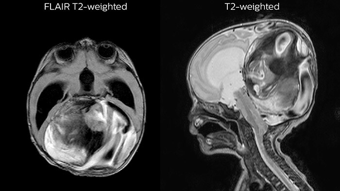
After three times of coiling and acryl based embolization
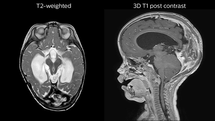
“We use this Compressed SENSE acceleration technology mainly for improving resolution without increasing scan time. We have already incorporated it into most of our ExamCards.”
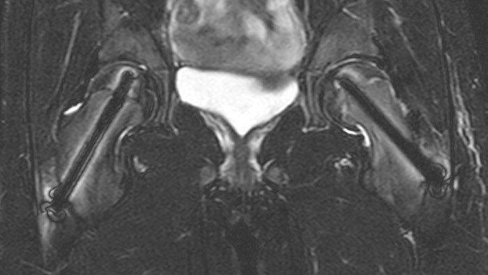
Slipped capital femoral epiphysis
After intervention with two cannulated titan screws, it is important to check that the circumference is normal and without necrosis. The screws can cause major metal artifacts, but O-MAR allows to improve visualization of tissue and bone in the near vicinity of MR Conditional orthopedic implants1. 1 Only for use with MR Safe or MR Conditional implants by strictly following the Instructions for Use
“We really appreciate how the Ambition allows us to change scan parameters quickly and easily; this is an extraordinary feature”
The need to adapt exams in real-time
The ability to perform comprehensive MRI scans tailored to every patient is vitally important for Dr. Junge and his team. The pediatric patients that arrive for an MRI are often not yet diagnosed, and the MRI exam is the first imaging they undergo.
“We may not know exactly what and where we need to scan beforehand,” says Dr. Junge. “During the examination – when seeing the first images – we may realize that we need to scan more anatomies or add different sequences in order to obtain all the relevant information immediately in the first exam. That is why we need flexibility during the exam. We constantly need to adapt our ExamCards as new clinical questions arise. We really appreciate how the Ambition allows us to change scan parameters quickly and easily; this is an extraordinary feature with Ambition, it’s extremely useful, I think.”
Robust ExamCards with flexibility to tailor
Having a collection of dedicated ExamCards is important, as these are the starting points of MRI examinations. “We begin every examination with one of the ExamCards from our collection,” says Dr. Junge. “Then during the examination, as new clinical questions arise based on the initial findings, we adjust and add sequences. This flexible way of working – ExamCards for the routine scans and the possibility to change a parameter with one click – I think that’s an great benefit of Philips MRI scanners.”
“Our application specialist initially helped us make our set of ExamCards for pediatric targets, which we built by adjusting the ExamCards from the standard set of Ambition. Each ExamCard bundles the sequences for a specific examination. Currently, we are still developing more ExamCards for the different age and size categories of our pediatric patients.”
"This flexible way of working – ExamCards for the routine scans and the possibility to change a parameter with one click – is an great benefit.”
Dedicated pediatric oncology ExamCards
An extensive set of dedicated pediatric ExamCards was developed in a collaboration with some expert users in Germany and based on the guideline from the European Society for Pediatric Oncology Brain Tumor Imaging Group (Nov 2017) and consensus of 11 German Philips MRI users (March 2019). Some highlights are:
Neuro-oncology ExamCards for 3.0T
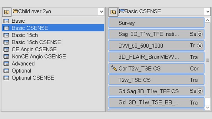
Body-oncology ExamCards for 1.5T
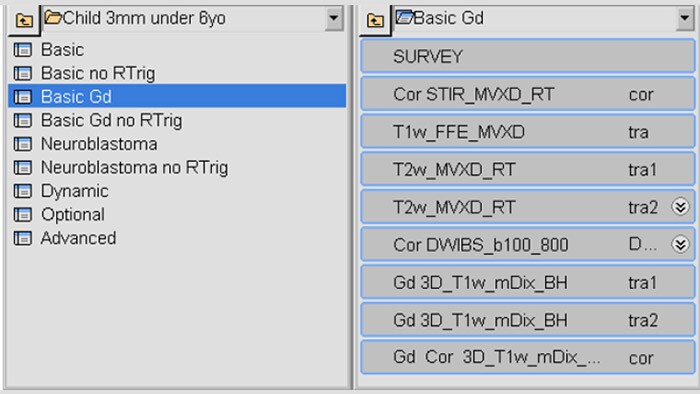
Examples of 3.0T neuro-oncology ExamCards (left) and 1.5T body-oncology ExamCards (right) for a selected age group. The list of sequences from the highlighted ExamCard is shown on the right. These ExamCards are part of the Philips DACH Pediatric Reference Scan Protocols
A leap in image quality thanks to progress in technology
“It was also a very pleasant surprise to us that we could get excellent resolution on other anatomies, as well, such as the spine, head, and knees, that we didn’t see previously. That is something that helps us tremendously as anatomy of children can be very small.”
The Ingenia Ambition gave Altona Children’s Hospital access to various novel scan techniques that are useful for pediatric imaging. “These modern technologies help us to scan anatomies where we saw challenges in the past, such as in the abdomen and thorax. With Ambition, we can perform high quality abdominal scans. As a result, the number of abdominal scans has increased substantially compared to our practice with our old system,” says Dr. Junge.
“We like to use the VitalEye feature in every abdominal examination. In my opinion it is an excellent trigger feature, and a big improvement from the respiratory belt we were using before,” says Dr. Junge.
Features that provide remarkable benefits to Dr. Junge include Compressed SENSE, which allows to elevate spatial resolution, signal and scan time. MultiVane XD and 3D VANE XD employ radial k-space sampling and help to mitigate motion artifacts and improve robustness for different contrasts and for all age groups. The mDIXON FFE and mDIXON TSE methods nicely address challenges in fat-free imaging and provide multiple contrast types from one single scan. The achievable large field of view (FOV), high resolution and flexible echo times are certainly a huge benefit in examining children.
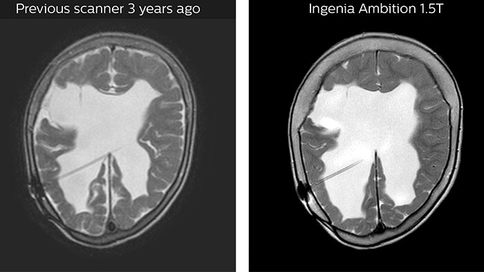
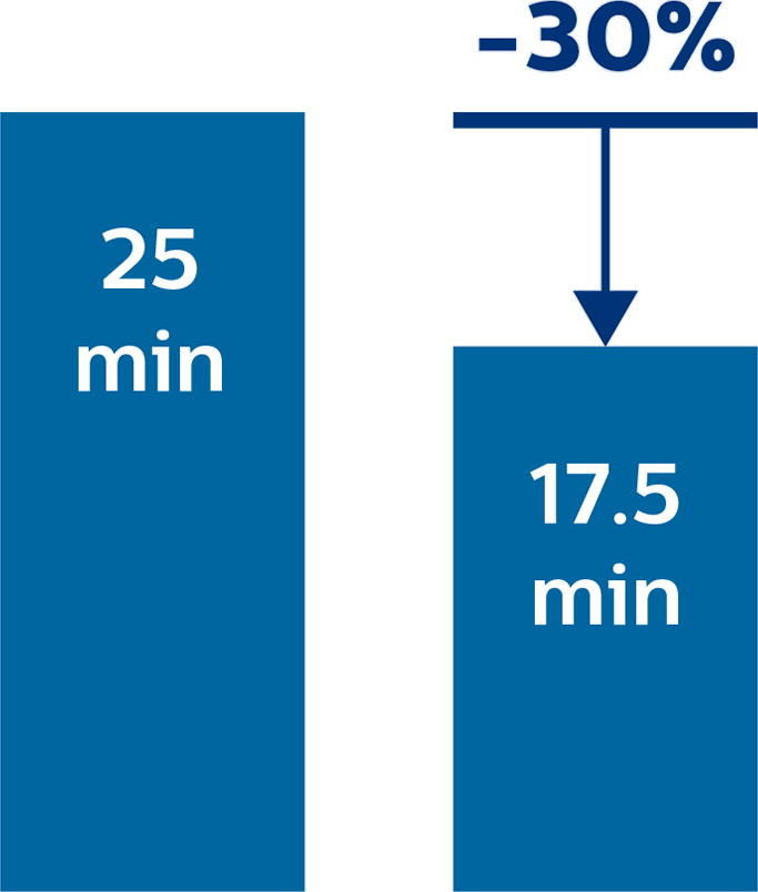
Hydrocephalus post hemorrhagic Both pictures show a ventriculoperitoneal shunt. With our previous scanner our hydrocephalus protocol needed about 25 min. With Ambition the examination time is about 14 min. including a CSF PCA sequence to show flow in the aqueduct.
Compressed SENSE
is an acceleration technology that allows to reduce scan time or to increase spatial resolution within the same scan time
Neuro oncology Basic ExamCard Sag 3D T1w TFE 3D FLAIR BrainVIEW aniso Sag 3D FLAIR BrainVIEW (optional) DWI b0 500 1000 Cor T2w_TSE T2w_TSE Sag 3D T1w TFE GD 3D T1w TSE GD
Survey
Compressed
SENSE
Compressed
SENSE
“We like to use VitalEye in every abdominal examination. In my opinion it is a big improvement from the respiratory belt we were using before.”
“Without much effort, we almost immediately doubled the number of patients per day”
Increasing the number of patients per day with Ambition
According to Dr. Junge, the department used to typically scan three or four patients per day with their previous system. However, this number instantly increased when they began using the Ambition. “Without much effort, we almost immediately doubled the number of patients to approximately six or eight per day,” says Dr. Junge. “We have only been using the system for three months and we plan to scan more patients in the future. I think our daily capacity can grow to ten or more patients per day.”

Altona Children’s Hospital
Reasons to buy turned into expectations that are met
“Our workflow benefits from the fact that our collection of dedicated ExamCards help us start a pediatric exam quickly and consistently. At the same time the scanner allows us to modify the parameters and sequences when we discover during the exam that a clinical question requires that. For patient satisfaction, the most impressive feature is the experience in the bore. That’s absolutely the highlight of the machine.” Dr. Junge also commented on the fully sealed magnet which contains only 7 liters of helium. “No helium can escape from the magnet, so no refills are needed, which represents a nice cost saving,” says Dr. Junge. The risk of a quench – with its associated disruptions and cost – is also eliminated.
Several factors contributed to the purchase of the Ingenia Ambition system. “Excellent image quality and high resolution have always been the most important criteria for us. It has been amazing for us to see the image resolution and consistent quality that we can obtain with Compressed SENSE throughout the body,” Dr. Junge says. “Also the mDIXON images are fantastic, and 3D VANE and other features as well.”
“It has been amazing for us to see the image resolution and quality that we can obtain with Compressed SENSE throughout the body. Also the mDIXON images are fantastic, and 3D VANE and other features as well.”
Meeting pediatric imaging goals with Ambition capabilities
“For a good diagnosis, we often need to look at multiple anatomies with several diagnostic possibilities and make adjustments during the scan. This can be challenging, but the Ambition provides us with great features, easy operation and high flexibility for obtaining high quality, comprehensive imaging in our pediatric patient population that covers a wide range of age, size and clinical indications. And besides, from day one, we also scan more patients than with our previous system.”
To conclude, Dr. Junge reiterates that the Ingenia Ambition with its unique magnet technology provides excellent scanning features as well as patient comfort. “Our young patients are surprised and fascinated by the Ambient room and the in-bore experience, which helps them undergo the examination,” he says. “This is key for successful pediatric examinations. However, most outstanding for me are the many features for obtaining high-end pictures with an 1.5T system.”
“The Ambition provides us with great features, easy operation and flexibility for obtaining high quality imaging in our pediatric patient population”
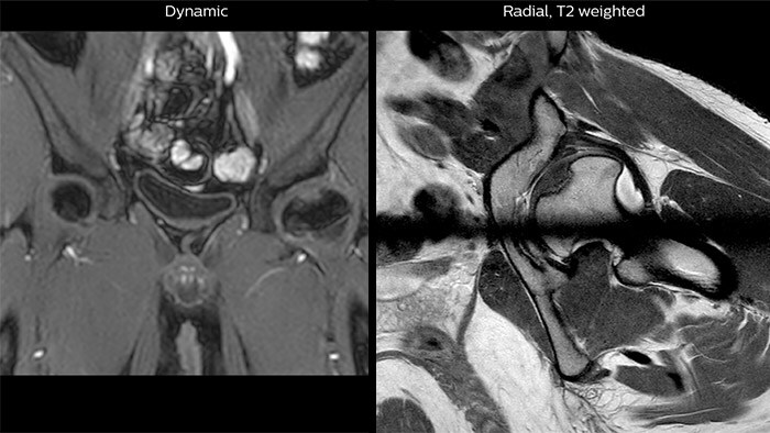
Perthes disease in left hip The affected area on the upper circumference of the left hip shows contrast uptake in the dynamic scan. The radial scan nicely depicts the hip area, despite the dark shape in the center that is inherent to the radial way of scanning.
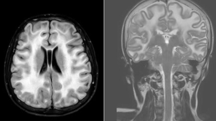
Leukodystrophy in a teenager
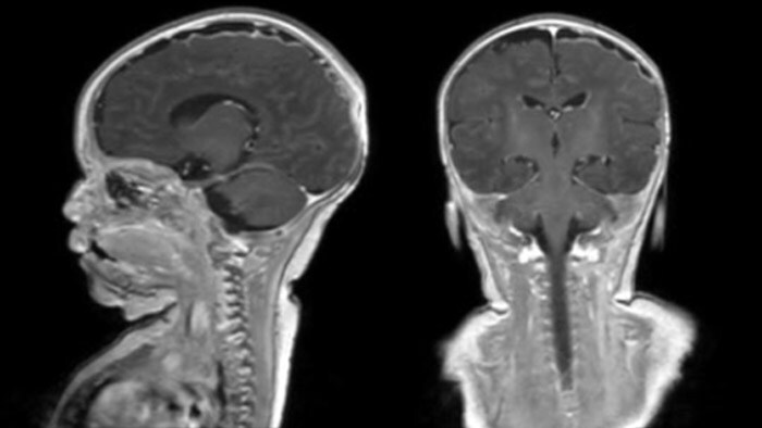
Rostral meningitis and arachnoiditis Both images are from the same 3D T1-weighted post contrast sequence in a newborn, under treatment.
Summary of experiences with Ingenia Ambition at Altona Children’s Hospital:
Results from case studies are not predictive of results in other cases. Results in other cases may vary.
Subscribe to FieldStrength
Our periodic FieldStrength MRI newsletter provides you articles on latest trends and insights, MRI best practices, clinical cases, application tips and more. Subscribe now to receive our free FieldStrength MRI newsletter via e-mail.
Stay in touch with Philips MRI
