
Tumor embolization
Highlighted products for this procedure
Resources
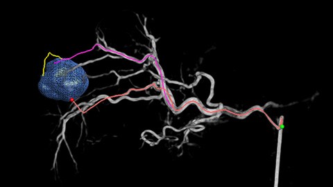
Automatic Feeder Detection with EmboGuide
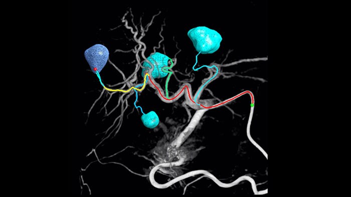
Adoption of chemo/radioembolization techniques such as TACE and SIRT drives the need for standardization and efficiency. Case after case, you must reliably and consistently locate the tumor(s), identify all feeder vessels, and plan/execute the appropriate interventional approach. Our Automatic Feeder Detection solution can significantly improve feeding artery detection compared to using Cone Beam CT alone. EmboGuide supports you in maximizing the efficacy of your TACE procedures as it potentially enhances your sensitivity, reduces false positives and maximizes inter-reader agreement.1
86%
57%
Less false Positive2
99.7%
Reader Agreement2
Tumor embolization
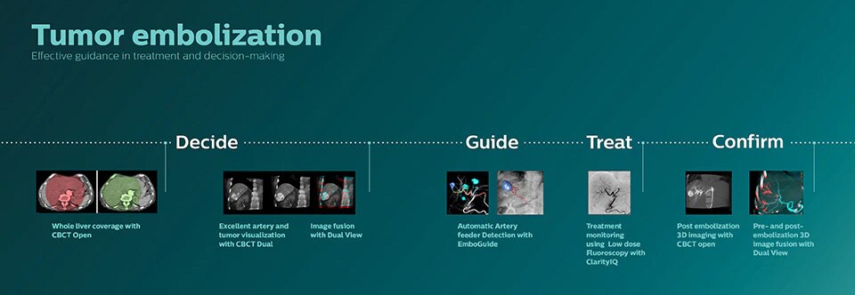
Effective guidance in treatment and decision making
The ability to detect and differentiate hepatic nodules and identify tiny feeder vessels is critical to determining proper therapy. Navigating to the region of interest by reaching all feeders, while remaining selective to the lesion, increases the opportunity for success. Confirmation of treatment endpoint and treatment success while the patient is still on the table boosts clinical outcome confidence.
Whole liver coverage with CBCT Open
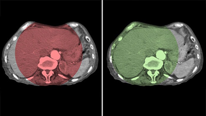
By opening the arc to the left of the patient, CBCT Open allows off center positioning of the patient table and therefore better centering of the FOV3-4. It significantly increases image coverage to help visualize tumors on the periphery of the liver.4
Optimizing artery and tumor visualization with CBCT Dual
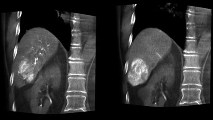
CBCT Dual enables 3D acquisition of an arterial phase to visualize vascular structures and a post-arterial (delayed phase) to visualize accumulation of contrast medium, in a single automatic step.5
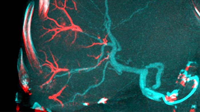
Image fusion with Dual View
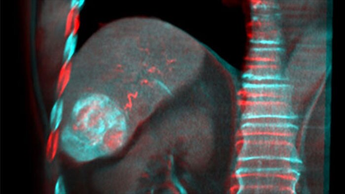
Dual View allows simultaneous visualization of two CBCT datasets. Both arterial and delayed phase can be displayed next to each other or in a single fused overlay view.
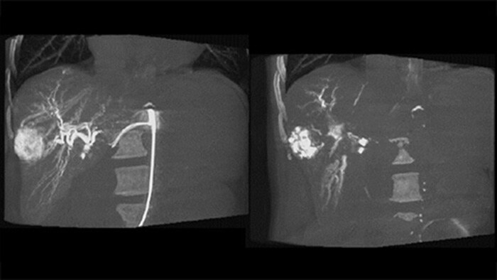
Post embolization 3D imaging with CBCT
A post embolization 3D acquisition allows you to visualize the targeted deposition of embolic material, such as Liopidol or radiopaque beads, in the tumor.5
Pre-and Post-embolization 3D image fusion with Dual View
Dual View allows simultaneous visualization of pre-embolization arterial phase 3D image and the post embolization image to assess treatment endpoint.
Explore the various procedures in interventional oncology and related clinical areas
-
![Tumor biopsy and ablation]()
Tumor biopsy and ablation
Solutions for tumor biopsies and ablations procedures offered by Philips image-guided therapy.
Click here to learn more -
![Prostate Artery Embolization (PAE)]()
Prostate Artery Embolization (PAE)
Effective guidance in treatment and decision making for prostate artery embolization.
Click here to learn more


Disclaimer Product availability is subject to country regulatory clearance. Please contact your local sales representative to check the availability in your country. References 1 Miyayama S, et al. Identification of small hepatocellular carcinoma and tumor-feeding branches with cone-beam CT guidance technology during transcatheter arterial chemoembolization. J Vasc Interv Radiol. 2013; 24(4):501-8. 2 Chiaradia et al, J J,Sensitivity and Reproducibility of AFD Software for HCC, Vasc Interv Radiol 2018;29:425-431. 3 Schernthaner RE et al, Feasibility of a Modified Cone-Beam CT Rotation Trajectory to Improve Liver Periphery Visualization during Transarterial Chemoembolization, Radiology. 2015; 277(3):833–4. 4 Loffroy R, et al. Comparing the detectability of hepatocellular carcinoma by C-arm dual-phase cone-beam computed tomography during hepatic arteriography with conventional contrast-enhanced magnetic resonance imaging. Cardiovasc Intervent Radiol. 2012;35(1):97-104. 5 Levi E.B. et al, First Human Experience with Directly Image-able Iodinated Embolization Microbeads, Cardiovascular and interventional radiology, vol 39, issue 8, 1177-1186, 2016